
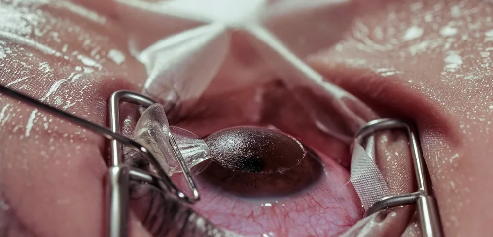
What is retinal detachment, and why does timing matter?
Retinal detachment happens when the retina lifts away from the back of the eye, cutting off its blood and oxygen supply. If they seek care fast, many people keep useful vision, but delays can reduce outcomes, especially if the macula is involved.
Australian eye clinics and hospital emergency departments treat suspected cases as time-critical, often same day.
What symptoms suggest they need urgent assessment in Australia?
They should treat sudden flashes, a rapid increase in floaters, or a “curtain” moving across vision as urgent warning signs. Blurred or distorted central vision may suggest macular involvement.
In Australia, they can present to an emergency department or contact an optometrist or ophthalmologist for same-day review, as prompt diagnosis can change which retinal detachment treatments are suitable.
How do clinicians decide which retinal detachment treatments are appropriate?
They decide based on whether it is a retinal tear only, a localised detachment, or a more extensive detachment. The presence of vitreous traction, bleeding, scar tissue (PVR), lens status, and whether the macula is still attached also matters.
Imaging and examination typically include dilated fundoscopy, widefield retinal photos, and often ocular ultrasound if the view is obscured.
When is laser used, and how does it differ from surgery?
Laser is usually used to “spot-weld” around a retinal tear or small hole before a detachment spreads. It is not the main treatment once a large section of retina has lifted.
In Australia, laser is commonly done in rooms under drops, and it is one of the least invasive retinal detachment treatments when the retina is not detached or only minimally threatened.
What is cryotherapy, and when might they receive it?
Cryotherapy uses a freezing probe on the outside of the eye to create a scar seal around a tear. It is helpful when laser access is difficult, such as with very peripheral tears or poor visibility.
They may receive cryotherapy as part of surgery or occasionally as a stand-alone sealing method, depending on the surgeon’s judgement and the tear’s position.
What is pneumatic retinopexy, and who is it best for?
Pneumatic retinopexy involves injecting a gas bubble into the eye so it presses the retina back into place while laser or cryotherapy seals the tear. It is generally suited to selected detachments with a single or small cluster of tears in the upper retina.
In Australia, it is less common than vitrectomy for broader detachments, but it remains a useful option among retinal detachment treatments when case selection is ideal and they can posture reliably.
What is vitrectomy, and why is it now so common?
Vitrectomy removes the vitreous gel that can tug on the retina, allowing the surgeon to flatten the retina and seal tears with laser. The eye is then filled with a gas bubble or silicone oil to keep the retina in place while healing occurs.
Modern small-gauge instruments have made vitrectomy faster and more precise, which is why it is a leading choice among retinal detachment treatments in Australian practice, especially for complex tears or traction.
What is scleral buckle surgery, and how does it differ from vitrectomy?
A scleral buckle places a silicone band around the eye to gently indent the wall, reducing traction and supporting the tear from the outside. It can be used alone or combined with vitrectomy.
In Australia, buckle surgery is often considered for younger patients, certain peripheral tear patterns, and some cases where preserving the vitreous is advantageous. It remains a key part of retinal detachment treatments, even if used less than vitrectomy overall.
How do surgeons choose between gas and silicone oil?
Gas is commonly used when they can follow posturing instructions and when the surgeon expects the retina to stay stable as the bubble slowly absorbs. Silicone oil may be used when longer-term internal support is needed, such as with significant scar tissue, giant tears, or when they cannot posture.
A practical Australian consideration is air travel: they cannot fly with an intraocular gas bubble due to dangerous eye pressure rise, which can influence retinal detachment treatments planning.

What does posturing involve, and why does it matter?
Posturing means holding the head in a specific position so the gas bubble presses on the retinal tear. The exact posture and duration depend on tear location and surgeon preference.
They may need to posture for days, sometimes longer, and adherence can affect results. Posturing requirements are a major way some retinal detachment treatments differ in real-life burden.
What can they expect on the day of treatment in Australia?
Many procedures occur in private day-surgery facilities or public hospital theatres, depending on urgency and availability. Patients may have local anaesthetic with sedation or a general anaesthetic, particularly for longer or more complex repairs, making retinal surgery anaesthesia options an important part of pre-operative planning.
Afterwards, they usually go home the same day with drops and activity limits, unless medical factors require observation.
How do recovery timelines differ between treatments?
Laser or cryotherapy alone often has a quicker functional recovery, although vision may still fluctuate for days. Vitrectomy or buckle repairs can take weeks to months for vision to stabilise, particularly if the macula was detached.
Among retinal detachment treatments, recovery is often more about retinal status and macular involvement than the surgical label, but the internal tamponade choice (gas vs oil) also changes the timeline.
What risks and side effects should they understand?
All procedures carry risks, including infection, bleeding, raised eye pressure, or recurrent detachment. Vitrectomy increases the likelihood of cataract progression in people who still have their natural lens.
They should also expect temporary blurred vision with gas, as the bubble blocks sight until it shrinks. Understanding these trade-offs helps them compare retinal detachment treatments realistically.
How successful are retinal detachment treatments in Australia?
Success is often discussed as anatomical reattachment after one operation, and many cases achieve this, though complex detachments may need more than one procedure. Visual recovery depends heavily on whether the macula detached and for how long.
Australian retinal surgeons typically quote outcomes tailored to the person’s retinal findings, not a single blanket percentage, because retinal detachment treatments success varies by complexity.
What happens if the detachment comes back?
A recurrent detachment can occur if new tears form, scar tissue develops, or the original break does not seal fully. They may need repeat vitrectomy, additional laser, a buckle, or silicone oil if not already used.
Follow-up is crucial in the first weeks and months, because early detection can make further retinal detachment treatments more straightforward.
How do costs and access work across Australia?
Access depends on location, surgeon availability, and whether they use the public system, private health insurance, or self-fund. Emergency treatment is prioritised medically, but timing and choice of facility can vary between metro and regional areas.
They can ask for written cost estimates in private care, including surgeon, anaesthetist, and hospital fees, as financial planning can influence which retinal detachment treatments pathway they pursue.
What questions should they ask their Australian eye surgeon?
They should ask which tear(s) were found, whether the macula is detached, and why one procedure is preferred over another. They can also ask about posturing time, tamponade choice, flying restrictions, expected vision, and follow-up timing.
Clear questions help them understand how the recommended retinal detachment treatments fit their specific retina, lifestyle, and recovery capacity.
How can they reduce risk after treatment?
They should follow drop schedules, posture exactly as instructed, and attend all reviews. They should avoid heavy lifting and high-impact activity until cleared, and seek urgent care for worsening vision, increasing pain, or new flashes and floaters.
For some people, monitoring the other eye matters too, as retinal tears can occur bilaterally, and prevention can reduce the need for further retinal detachment treatments.
What is the key takeaway on how these treatments differ?
They differ mainly in invasiveness, where the repair is performed, whether it is approached from inside or outside the eye, and the level of aftercare required. Laser and cryotherapy can seal tears, gas-bubble approaches rely on positioning, while vitrectomy or buckle surgery can physically reattach and support the retina in larger or more complex detachments, making retinal damage symptoms important to recognise early.
In Australia, prompt assessment is the deciding factor that keeps more retinal detachment treatments on the table and improves the chance of preserving vision.

FAQs (Frequently Asked Questions)
What is retinal detachment and why is urgent treatment important?
Retinal detachment occurs when the retina lifts away from the back of the eye, cutting off its blood and oxygen supply. Prompt treatment is critical because delays can lead to permanent vision loss, especially if the macula—the central part of the retina—is involved. In Australia, suspected cases are treated as time-critical emergencies, often requiring same-day intervention to preserve useful vision.
What symptoms indicate the need for urgent retinal detachment assessment in Australia?
Sudden flashes of light, a rapid increase in floaters, or a shadow or “curtain” moving across vision are urgent warning signs. Blurred or distorted central vision may suggest macular involvement. Individuals experiencing these symptoms should seek immediate assessment at an Australian emergency department or contact an optometrist or ophthalmologist for same-day review to determine appropriate retinal detachment treatments.
How do clinicians decide which retinal detachment treatments are suitable?
Treatment choice depends on factors such as whether there is a retinal tear only, a localized detachment, or a more extensive detachment. Other considerations include vitreous traction presence, bleeding, scar tissue (proliferative vitreoretinopathy), lens status, and whether the macula remains attached. Examination typically involves dilated fundoscopy, widefield retinal imaging, and ocular ultrasound if needed to guide appropriate treatment selection.
What are the differences between laser treatment and surgery for retinal detachment?
Laser treatment is used to ‘spot-weld’ around small retinal tears or holes to prevent detachment spread and is minimally invasive, typically performed in-clinic under eye drops. Surgery becomes necessary when larger sections of retina have detached. Surgical options in Australia include vitrectomy and scleral buckle procedures that physically repair and reattach the retina.
What is pneumatic retinopexy and who is it best suited for?
Pneumatic retinopexy involves injecting a gas bubble into the eye to press the retina back into place while laser or cryotherapy seals the tear. It is best suited for selected detachments with single or small clusters of tears located in the upper retina. Although less common than vitrectomy in Australia for broader detachments, it remains a valuable option when patient selection and posturing compliance are ideal.
How do surgeons choose between using gas bubbles and silicone oil during retinal detachment surgery?
Gas bubbles are commonly used when patients can reliably maintain required head positioning (posturing) and when gradual absorption aligns with healing expectations. Silicone oil provides longer-term internal support and is preferred in cases with significant scar tissue, giant tears, or when posturing is not feasible. In Australia, considerations such as air travel restrictions—patients cannot fly with intraocular gas due to pressure risks—also influence this decision among retinal detachment treatments.